Pelvic Exam & Medical History
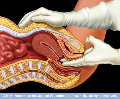
Some fibroids can be felt as lumps during a pelvic exam. During a pelvic exam, the doctor will also check for pregnancy-related conditions and other conditions, such as ovarian cysts. The doctor will also ask you about your medical history, particularly as it relates to menstrual bleeding patterns. Other causes of abnormal uterine bleeding and must also be considered.
Ultrasound
Ultrasound is the standard imaging technique for detecting uterine fibroids. The doctor will order transabdominal and transvaginal ultrasounds. Ultrasound is a painless technique, which uses sound waves to image the uterus and ovaries. In transabdominal ultrasound, the ultrasound probe is moved over the abdominal area. In transvaginal ultrasound, the probe is inserted into the vagina.
A variation of ultrasound, called hysterosonography, uses ultrasound along with saline (salt water) infused into the uterus to enhance the visualization of the uterus.
Hysteroscopy
Hysteroscopy is a procedure that may be used to detect the presence of fibroids, polyps, or other causes of bleeding. (It may also be used during surgical procedures to remove fibroids.) Hysteroscopy can be performed in a doctor’s office or in a hospital setting. The procedure uses a long flexible or rigid tube called a hysteroscope, which is inserted into the vagina and through the cervix to reach the uterus. A fiber optic light source and a tiny camera in the tube allow the doctor to view the cavity. The uterus is filled with saline or carbon dioxide to inflate the cavity and provide better viewing. This can cause cramping. Hysteroscopy is non-invasive and does not require incisions; however, a third of women report severe pain with the procedure. Local, regional, or general anesthesia may be given.
Laparoscopy
In some cases, laparoscopic surgery may be performed as a diagnostic procedure. Laparoscopy involves inserting a scope into a small incision made near the navel. Whereas hysteroscopy allows the doctor to view inside the uterus, laparoscopy provides a view of the outside of the uterus, including the ovaries, fallopian tubes, and general pelvic area.
Magnetic Resonance Imaging or MRI
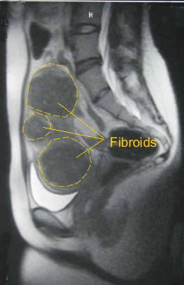
In some cases, MRI may be used in addition to ultrasound to evaluate women with fibroids. MRI is sometimes better able to delineate the location of a myoma or differentiate a myoma from adenomyosis. Women undergoing uterine fibroid embolization will undergo MRI prior to and after the procedure. Some women undergoing myomectomy will undergo MRI to better visualize their fibroids prior to surgery. Magnetic resonance imaging (MRI) is a noninvasive medical test that helps physicians diagnose and treat medical conditions.
MR imaging uses a powerful magnetic field, radio frequency pulses and a computer to produce detailed pictures of organs, soft tissues, bone and virtually all other internal body structures. The images can then be examined on a computer monitor, printed or copied to CD. MRI does not use ionizing radiation (x-rays). Detailed MR images allow physicians to better evaluate various parts of the body and certain diseases that may not be assessed adequately with other imaging methods such as x-ray, ultrasound or computed tomography (also called CT or CAT scanning).
Other Tests
In certain cases, the doctor may perform an endometrial biopsy to determine if there are abnormal cells in the lining of the uterus that suggest cancer. Endometrial biopsy can be performed in a doctor’s office, with or without anesthesia. Dilation & curettage (D&C) is a more invasive procedure that involves scraping the inside lining of the uterus. It can be used to take tissue samples and also as a procedure to help temporarily reduce heavy menstrual bleeding.
The doctor may also order a complete blood count (CBC) to check for signs of anemia.